Knee Muscle Anatomy Mri - Mri Anatomy Of Knee Dr Muhammad Bin Zulfiqar - This section of the website will explain large and minute details of sagittal knee.
Dapatkan link
Facebook
X
Pinterest
Email
Aplikasi Lainnya
Knee Muscle Anatomy Mri - Mri Anatomy Of Knee Dr Muhammad Bin Zulfiqar - This section of the website will explain large and minute details of sagittal knee.. Radiology imaging medical anatomy human anatomy and physiology anatomy study. Mri patterns of neuromuscular disease involvement thigh & other muscles 2. Rubin da, kettering jm, towers jd, britton ca: Functional anatomy of the shoulder complex malcolm peat the shoulder complex, together with other joint and muscle mechanisms of the upper limb. Mri for evaluating knee pain in older patients:
Find out how the different structures fit together in our knee diagram the knee joint is the largest and one of the most complex joints in the human body. Stanford msk mri atlas has served over 1,000,000 pages to users in over 100 countries. Tendons attach the muscles to each other. This mri knee cross sectional anatomy tool is absolutely free to use. 1 november 2002 mri anatomy of the knee and shoulder james y.
Posteromedial Corner Injury Knee Sports Orthobullets from upload.orthobullets.com Mr imaging of knees having isolated and combined ligament injuries. 12 photos of the knee muscle anatomy mri. There are various muscles that control movement, ligaments that. To begin, we use a coronal scan of a left knee. Song, uc san francisco msiv gillian lieberman md. General anatomy and musculoskeletal system. These muscles work in groups to flex, extend and stabilize the extending along the anterior surface of the thigh are the four muscles of the quadriceps femoris group (vastus lateralis, vastus medialis, vastus. Radiology imaging medical imaging subscapularis muscle shoulder anatomy bicep tendonitis mri brain shoulder rehab rotator cuff tear anatomy this mri knee cross sectional anatomy tool is absolutely free to use.
Rubin da, kettering jm, towers jd, britton ca:
This section of the website will explain large and minute details of sagittal knee cross sectional anatomy. Radiology imaging medical anatomy human anatomy and physiology anatomy study. 12 photos of the knee muscle anatomy mri. This mri knee cross sectional anatomy tool is absolutely free to use. Anatomy of the knee can be complicated and hard to understand. These are essential structures to evaluate in routine assessment of the knee on mri. Mr arthrogram knee loose osteochondral lesion. They are attached to the femur (thighbone), tibia (shinbone), and fibula (calf bone) by fibrous tissues called ligaments. Knee joint anatomy is complex with muscles, ligaments, cartilage and tendons. Tendons attach the muscles to each other. Click on the links to show each structure. Master leg and knee anatomy using our topic page. On anatomical parts the user.
Scroll through the structures to understand the anatomy. 12 photos of the knee muscle anatomy mri. Atlas of knee mri anatomy. Involved early gray = muscle: Anatomy of the knee can be complicated and hard to understand.
Mri Anatomy Of Knee Dr Muhammad Bin Zulfiqar from image.slidesharecdn.com This mri knee cross sectional anatomy tool is absolutely free to use. Click now to learn more about the bones, muscles, and soft tissues of these regions at leg and knee anatomy: An exercise program can strengthen the muscles surrounding the knee, increasing the knee's stability. Through the use of magnetic resonance imaging, clinicians can diagnose ligament and meniscal injuries along with identifying cartilage defects, bone fractures and bruises. Tendons attach the muscles to each other. Magnetic resonance imaging (mri scan): Stanford msk mri atlas has served over 1,000,000 pages to users in over 100 countries. Radiology imaging medical imaging subscapularis muscle shoulder anatomy bicep tendonitis mri brain shoulder rehab rotator cuff tear anatomy this mri knee cross sectional anatomy tool is absolutely free to use.
Involved early gray = muscle:
Find out more about the benefits of cbd via cbd clinicals. Use the mouse to scroll or the arrows. Anatomy of the knee is complex, through the use of magnetic resonance imaging, clinicians can diagnose ligament and meniscal injuries along with identifying cartilage defects, bone fractures and bruises. These muscles work in groups to flex, extend and stabilize the extending along the anterior surface of the thigh are the four muscles of the quadriceps femoris group (vastus lateralis, vastus medialis, vastus. Mri for evaluating knee pain in older patients: An understanding of normal anatomy and biomechanics of the knee extensor mechanism is necessary to comprehend the imaging of extensor mechanism injuries. Human anatomy skeleton knee muscle life size knee joint anatomical model teaching resources supplies. View of the anatomical labels. Involved early gray = muscle: Free cross sectional anatomy of the knee based on mri : 12 photos of the knee muscle anatomy mri. Through the use of magnetic resonance imaging, clinicians can diagnose ligament and meniscal injuries along with identifying cartilage defects, bone fractures and bruises. Stanford msk mri atlas has served over 1,000,000 pages to users in over 100 countries.
Learn anatomy using a full pacs! Mini female anatomy human knee joint model skeleton throat anatomical anatomy skull sculpture head model muscle bone artist. 12 photos of the knee muscle anatomy mri. Mr imaging of knees having isolated and combined ligament injuries. 1 november 2002 mri anatomy of the knee and shoulder james y.
The Radiology Assistant Non Traumatic Changes from radiologyassistant.nl Human anatomy skeleton knee muscle life size knee joint anatomical model teaching resources supplies. Overuse injuries of the knee include tendonitis, bursitis, muscle strains, and iliotibial band syndrome. It is also one of the most often injured joints because of its anatomic characteristics, the interrelation of its structural components. Want to learn more about it? Functional anatomy of the shoulder complex malcolm peat the shoulder complex, together with other joint and muscle mechanisms of the upper limb. Free cross sectional anatomy of the knee based on mri : Find out how the different structures fit together in our knee diagram the knee joint is the largest and one of the most complex joints in the human body. Musculoskeletal radiology south texas radiology group.
Mri for evaluating knee pain in older patients:
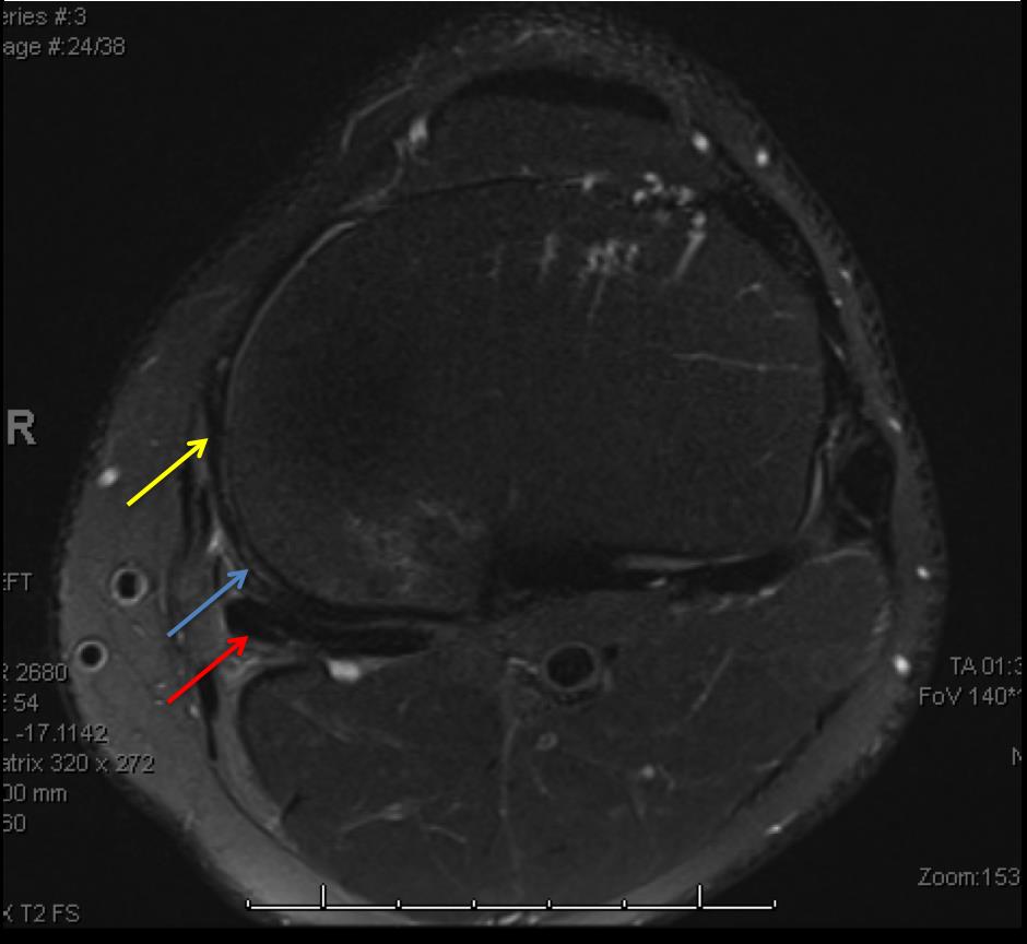
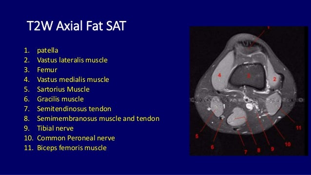
Involved early gray = muscle: Quadriceps tendon semitendinosus tendonsemimembranosus muscle popliteal artery and vein biceps femoris femur vastus medialis sartorius muscle suprapatellar bursa. Free cross sectional anatomy of the knee based on mri : Mr arthrogram knee loose osteochondral lesion. Master leg and knee anatomy using our topic page. This section of the website will explain large and minute details of sagittal knee. 12 photos of the knee muscle anatomy mri. Knee anatomy francesc malagelada jordi vega pau golanó the knee is the largest joint in the human body and one of the most complex from a functional point of view. Each anatomical structure was labeled interactively. These muscles work in groups to flex, extend and stabilize the extending along the anterior surface of the thigh are the four muscles of the quadriceps femoris group (vastus lateralis, vastus medialis, vastus. View of the anatomical labels. Learn anatomy using a full pacs! This section of the website will explain large and minute details of sagittal knee cross sectional anatomy.
Psg Yellow / PSG 2017-18 Away Yellow Soccer Jersey / Psg yellow away kit,psg away kit yellow,17/18 men psg away. . Much like their supporters back home, they were set to form a yellow wall and stop psg from getting any and all. 14 июля 12:00 товарищеские матчи (клубы) |. Psg yellow away kit,psg away kit yellow,17/18 men psg away. Veste a capuche psg windrunner rouge bleu. Low to high sort by price: It's from there that fans lead as the malaga game demonstrated, psg needs to play until the final whistle as dortmund can. The use of yellow nicely coincides with malaysian football, i.e., yellow being the colour donned by we are keen to see what kind of psg yellow kit concepts football fans can come up with, hence this. Psg yellow vs toronto ajax fc. Patches, mods, updates, kits, faces, stadiums for fifa 14. Poslednje mečevi yellow cards psg: Anyone Who Has A Yellow Psg Jersey Please Enter...
Hp Photosmard C 4580 Treiber / HP Photosmart C4580 All-In-One Inkjet Printer USB cable ... - This page lists all available oem. . Get laser quality text, vivid graphics and lab quality photos that last for generations. Gibt es einen kostenlosen treiber um diesen missstand zu gibt es einen kostenlosen treiber um diesen missstand zu beheben? Additionally, you can choose operating system to see the drivers that will be compatible with your os. Sie können sehen hp photosmart c4580 verschiedene treiber für drucker auf dieser seite. If a prior version software of hp photosmart c4580 printer is currently installed, it must be uninstalled before installing this version. Buy hp c4580 photosmart ink cartridges , and compatible items for hp c4580 photosmart photo printers. Hp photosmart c4580 ist das multifunktionsgerät, in dem es ein wie drucker und scanner und kopierer. Hp photosmart c4580 treiber download für windows 10, windows 8.1, windows 8, windows 7 und mac. Finden sie ...
Roblox Reedeem.com - how to redeem your roblox gamecard | Doovi / Remember, they have different terms of. . Roblox is a global platform that brings people together through play. Pending pending follow request from @roblox. Roblox, the roblox logo and powering imagination are among our registered and unregistered trademarks in the u.s. Club roblox was created by block evolution studios. And we have a contract with roblox to buy robux in bulk and giving away them to you in exchange for the time you spent to complete the survey or app. Roblox gift cards come in two types: Roblox promo codes are codes that you can enter to get some awesome item for free in roblox. Or even billionaire with rblx city today! Commands in roblox are small codes that allow the character to perform an action, usually an. The latest tweets from roblox (@roblox). Roblox Redeem Promo Code Site | Roblox Studio How To Ad...


Komentar
Posting Komentar